Decloitre (1963) recognizes 5 species of Arcella in Venezuela. As far as I know, there are no other species checklists of testate amoebas of Venezuela. Decloitre (1963) listed a total of 76 species of testate amoebas species for Venezuela in 15 different genera. The following is the list of species for the genus Arcella.
Genus: Arcella Ehrenberg
Arcella conica Ehrenberg
Arcella hemisphaerica Perty
Arcella hemisphaerica var gibba Deflandre
Arcella lobostoma Deflandre
Arcella polypora Pénard
Decloitre, L. 1963. Rhizopodes Thécamoebiens du Vénézuéla. Proceedings of the Zoological Society of London 141(3): 497-514.
Thursday, December 21, 2006
Arcella sp4


Specimen #25312-313:
Diameter shell: 100.8 µm
Diameter aperture: 36.5 µm
Height shell: 157.4 µm
Bucal depth: 9.2 µm
At first I believed this specimen to be Arcella mitrata var pyriformis but if you see on pic 25313, there is no crenulations on the aperture. Unfortunately I have no pictures for the apical view.
Arcella sp3


Specimen #25310-311:
Diameter shell: 160.3 µm
Diameter aperture: 48.7 µm
This specimen is similar to Arcella vulgaris var. undulata but the apreture is crenulated.
Arcella vulgaris f. undulata? Deflandre


Specimen #13854-856:
Diameter shell: 117.4 µm
Diameter aperture: 23.9 µm
Specimen #24962-963:
Diameter shell: 151.0 µm
Diameter aperture: 28.3 µm
Arcella mitrata? Leidy


Specimen # 24973-974:
Diameter shell: 113.2 µm
Diameter aperture: 32.5 µm
Height shell: 115.8 µm
Bucal depth: 13.1 µm
Specimen #24975:
Diameter shell: 115.7 µm
Diameter aperture: 37.4 µm
Height shell: 127.2 µm
Bucal depth: 13.4 µm
Specimen #24971:
Diameter shell: 114.2 µm
Diameter aperture: 33.4 µm
Height shell: 117.4 µm
Bucal depth: 13.7 µm
Arcella mitrata cf pyriformis? Deflandre


Specimen # 25331:
Diameter shell: 125.8 µm
Diameter aperture: 40.5 µm
Height shell: 135.6 µm
Bucal depth: 13.2 µm
Specimen # 25316:
Diameter shell: 121.4 µm
Diameter aperture: 42.7 µm
Height shell: 134.3 µm
Bucal depth: 9.6 µm
This two specimens seem to be Arcella mitrata var pyriformis but they are not tall enough. It seems to be an intermediate form between A. mitrata and A. mitrata var pyriformis.
Tuesday, December 19, 2006
Arcella cf. mitrata var pyriformis? Deflandre


Specimen #25220-221:
Diameter shell: 112.5 µm
Diameter aperture: 40.5 µm
Height shell: 147.1 µm
Bucal depth: 15.4 µm
This specimen looks like Arcella mitrata var pyriformis Deflandre 1928. Unfortunately, I do not have a picture showing the apical view.
Arcella gibbosa var levis? Deflandre

Specimen #25517:
Diameter shell: 93.5 µm
Diameter aperture: 37.2 µm
Height shell 68.1 µm
Bucal depth: 12.1 µm
Diameter shell: 93.5 µm
Diameter aperture: 37.2 µm
Height shell 68.1 µm
Bucal depth: 12.1 µm
Arcella costata var angulosa? (Perty) Playfair


Specimen #25356-358:
Diameter shell: 110.3 µm
Diameter aperture: 32.3 µm
Height shell: 68.4 µm
Bucal depth: 24.1 µm
Arcella costata Ehrenberg


Specimen #25602-604 (left):
Diameter shell: 108.0 µm
Diameter aperture: 37.4 µm
Height shell: 57.0 µm
Specimen #25602-604 (right):
Diameter shell: 105.8 µm
Diameter aperture: 38.1 µm
Arcella sp2


Specimen # 22187:
Diameter shell: 91.4 µm
Specimen # 21892:
Diameter shell: 95.5 µm
Diameter shell: 95.5 µm
Diameter aperture: 24.5 µm
Specimen # 20577:
Diameter shell: 112.9 µm
Specimen # 20598:
Diameter shell: 104.1 µm
Diameter shell: 104.1 µm
I could not measure the diameter of the aperture for some of the specimens. Unfortunately, all these specimens were only photographed on one side (apical view). For now on I will try to photograph all the necessary diagnostic taxonomic characters and all the possible views.
I think all these specimens are similar to Arcella costata. I posted Specimen #20577 earlier. It appears in a picture next to another specimen that I called Arcella megastoma.
Arcella sp1


Diameter shell: 100.8 µm
Diameter aperture: 33.6 µm
These three pictures were taken on the same specimen. I did not know how important is to take pictures of all sides of testate amoebas. It is a shame that I have only pictures showing the apical views of a lot of testate amoebas. I started taken pictures of all sides of these organisms a month ago. The three pictures I am showing here were one of my first attempts to have a complete series of all sides of any specimen. Although these pictures are not very good (I failed placing testate amoebas in the right position) at least they could be helpful for a preliminar identification.
In this specimen, I can barely see a crenulated aperture, but because the shape of the test, I could not say anything conclusive about its identification. This specimen is similar to Arcella leidyana Deflandre 1928 but with a smaller and less tall test.
Arcella crenulata? Deflandre


Specimen #21380:
Diameter shell: 100.2 µm
Diameter aperture: 32.3 µm
Specimen #7386:
Diameter shell: 109.5 µm
Diameter aperture: 24.9 µm
Specimen #12094:
Diameter shell: 143.3 µm
Diameter aperture: 26.5 µm
Arcella costata? Ehrenberg


Specimen #25054:
Diameter shell: 101.1 µm
Specimen #22491:
Diameter shell: 108.8 µm
Diameter aperture: 30.5 µm
Specimen #21041:
Diameter shell: 97.6 µm
Diameter aperture: 29.9 µm
Specimen #10864:
Diameter shell: 121.8 µm
Diameter aperture: 34.6 µm

Arcella megastoma Pénard & Arcella costata? Ehrenberg

Arcella megastoma (right)
Diameter aperture: 351.2 µm
Diameter shell: 171.1 µm
Arcella costata? (left)
Diameter aperture: 112.9 µm
Arcella megastoma Pénard


Specimen #10981-982:
Diameter of shell: 255.4 µm
Diameter of aperture: 125.3 µm
Monday, December 4, 2006

Thursday, November 30, 2006
Rio Cinaruco pictures
This series of pictures will allow you to see how the river looks. Most of the pictures were taken during 2002-2003 field seasons. Field work done during that time was supported by a NSF grant to K. Winemiller (TAMU), D. Roelke (TAMU) and J. Cotner (U. Minnesota) to study different aspects of benthic ecology in the Rio Cinaruco. More information on that NSF project can be obtained from the webpage of Kirk Winemiller at TAMU. Otherwise stated, all pictures published here were taken by Jose Montoya.
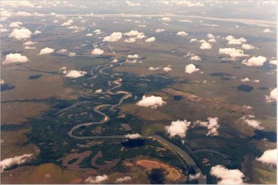
Rio Cinaruco aereal view. Sometime during the dry season of 1998. This picture shows the extent of geomorphological heterogeneity on this segment of the river. Note the different types of lakes and the sandy beaches on the main channel of the Cinaruco. The Orinoco is shown on the upper part of the picture.
Photo taken by María Mercedes Castillo (Universidad Simón Bolívar, Caracas, Venezuela)
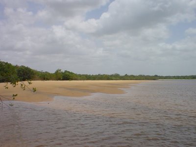
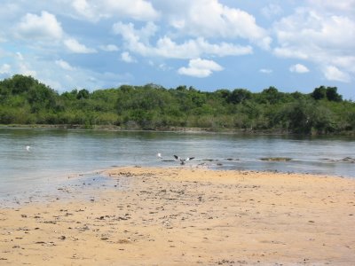 Sandy beach at Rio Cinaruco.
 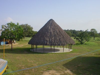 Field camp at Rio Cinaruco during March 2003 (Dry season, low-water period).
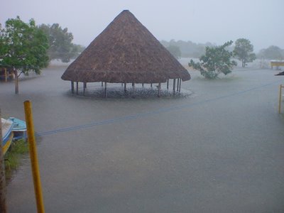
Field camp at Rio Cinaruco during late June 2003 (Wet season, high-water period).
 Rio Cinaruco sandy beaches in early May 2003 (rising-water period).
Subscribe to:
Comments (Atom)


